
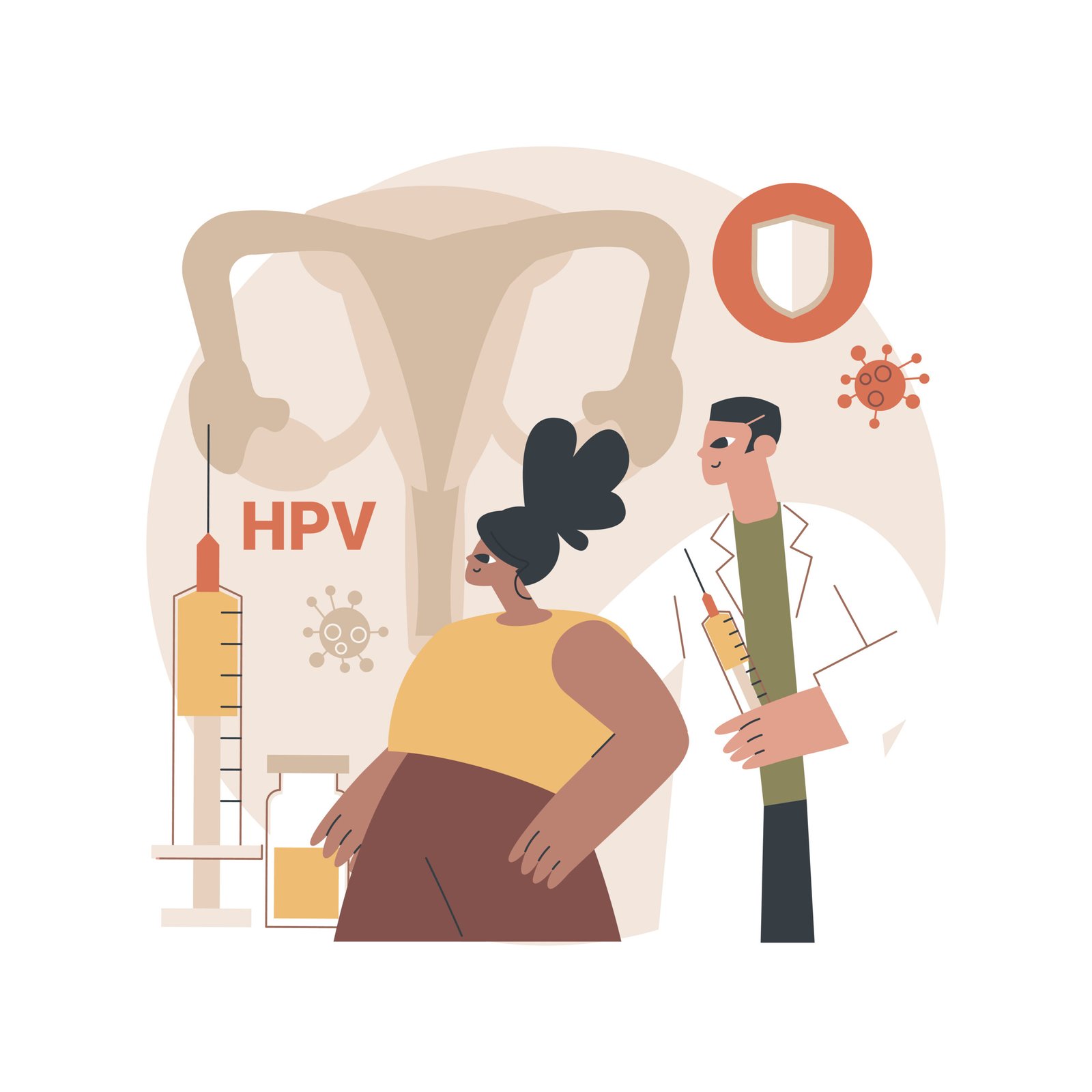
Cervical cancer, which is mostly caused by the human papillomavirus (HPV), is largely preventable through vaccination. Early detection through screening methods can help identify the HPV virus early. Early detection greatly improves the chances of successful treatment and can prevent any early cervical cell changes from becoming cancer. Also it is important to be aware of any signs and symptoms of cervical cancer which can help avoid unnecessary delays in diagnosis.
Let’s understand screening and its methods for detecting cervical cancer early
Cervical cancer screening helps find early signs of the disease, like precancerous lesions (abnormal cells in the cervix that can become cancerous) and HPV infections (a virus that can cause these abnormal changes in cervical cells), allowing for easier treatment and prevention of cervical cancer development or spread.
Below are the different methods of screening for early detection of cervical cancer
VISUAL INSPECTION WITH ACETIC ACID (VIA)

Visual inspection with acetic acid is a test to detect precancerous cervical lesions.This test requires short time, is conducted in privacy and is a painless procedure. For conducting this test, you need to lie down on the examination table, after removing your undergarments.
The examining health personnel after wearing gloves will insert a speculum inside your vagina and locate your cervix.The health personnel will then apply 5% acetic acid solution on your cervix using a cotton swab. After 2-3 mins, Lugol’s Iodine solution will be applied to your cervix. The doctor shall observe changes and remove the speculum. The VIA test is done.
Results are provided to women as VIA positive, which indicates a precancerous lesion, or VIA negative which indicates no lesion.
HUMAN PAPILLOMA VIRUS (HPV) TEST
In this test, after a tool called a speculum is gently put into the vagina to see the cervix, a special brush collects a small sample of cells from the cervix. This sample is then placed in a bottle with a liquid solution and sent to a lab for testing. The test helps find infections caused by HPV, a virus that can lead to cervical cancer.
This screening method is crucial for detecting cervical cancer before any symptoms appear, allowing for timely intervention to prevent its development.
PAP TEST OR PAP SMEAR

In this test, a cotton-tipped stick or brush gently collects cells from the cervix, which is the lower part of the uterus. These cells are then placed on a glass slide and examined under a microscope by a doctor or a lab technician.
This helps to check for any abnormalities or changes in the cells that could indicate a risk of cervical cancer. A Pap test also sometimes finds conditions that are not cancer, such as infection or inflammation.
If the result is positive for any Screening test it does not mean you have cancer. It means you may need to undergo further tests which are described below.
Biopsy: If cervical pre cancer is suspected, a small sample of the suspicious area is taken for examination.
Endocervical Curettage: If pre cancer is suspected in the inner part of the cervix, a small sample is scraped and examined.
Colposcopy: A procedure to closely examine the cervix for any abnormal areas.

In the majority of cases, test results will be normal, indicating a healthy cervix. Women with normal results are recommended to have a test again in 3-5 years’ time, which may vary according to country guidelines.
It is important for all women, even if their test result is normal, to take another test in the future, since changes in the cervical cells can develop at any point in time.
With HPV tests, if the HPV test result is negative (that is, no HPV infection is detected) and the woman is over 30 years of age, there is a lower chance for cervical cancer to develop. In this case, women are recommended to have an HPV test again in 5-10 years.

- Cancer Research UK / Cervical Cancer
- Cancer Research UK / Cervical Cancer / Getting diagnosed with cervical cancer
- Cancer Research UK / Cervical Cancer / Getting diagnosed with cervical cancer / Cervical screening
- Tata Medical Center / Preventive Oncology
- American Cancer Society / All About Cancer
- American Cancer Society / All About Cancer / Cervical Cancer
- American Cancer Society / All About Cancer / Cervical Cancer / Cervical Cancer Early Detection, Diagnosis, and Staging
- A Training Module on Cancer Awareness for Prevention and Early Detection of Breast and Uterine Cervix Cancer
- American Cancer Society / The Pap (Papanicolaou) Test