
Early detection of oral cancer through screening is crucial for successful treatment and improving recovery chances. Screening helps catch early signs and symptoms of oral cancer.
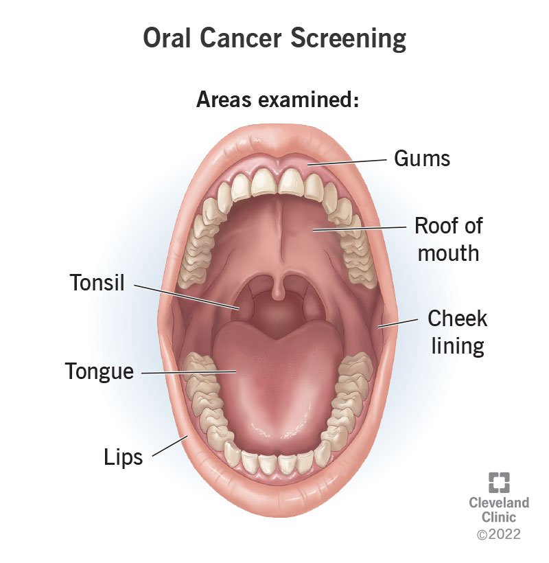
These screenings involve thorough clinical examinations of the mouth, including the lips, gums, tongue, and lining of the cheeks by dentists and healthcare professionals who are well-trained.
Additionally, individuals can perform oral self-examinations at home in a step-by-step manner for their own health awareness. If they suspect any changes that might be cancerous, individuals can visit a dentist early before the cancer reaches a severe stage.
It is important to remember that not every sign and change can be attributed to cancer; therefore, it plays a crucial role to get checked by healthcare professionals regularly.
Thus, regular clinical examinations and self-examinations at home are key for early oral cancer detection, helping to minimize the severity of treatment required
Let’s understand the two types of examinations done as a part of screening for finding oral cancer early
Clinical Examination
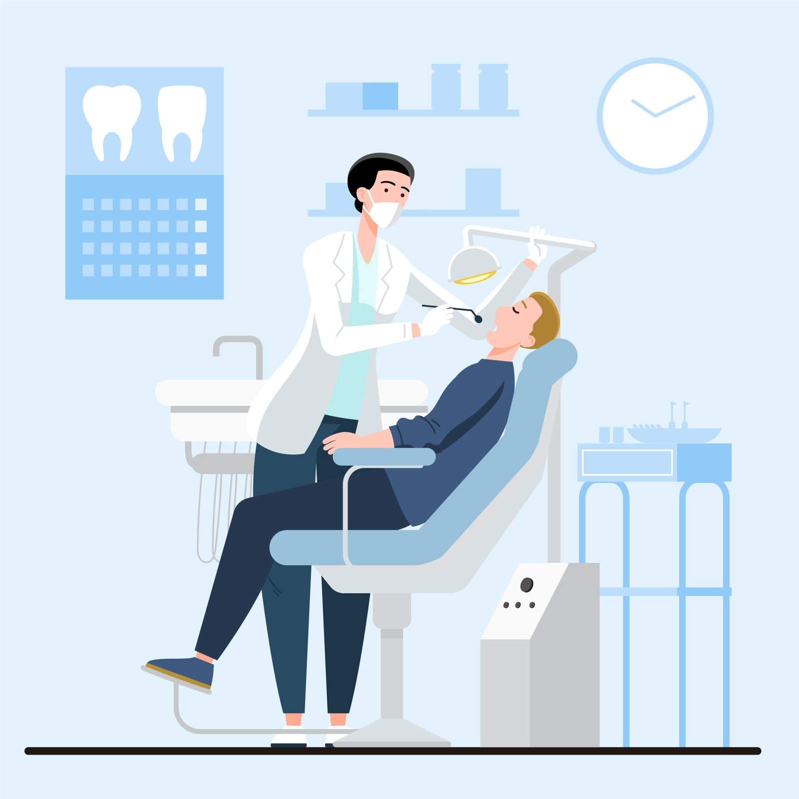
For conducting a clinical examination, also known as clinical oral visual inspection, your dentist or healthcare provider may use a combination of oral cancer screening methods, including a visual exam, palpation, and oral screening dyes and lights. They may also take photos of any abnormal areas to monitor them.
Self Examination

Oral self-examination means looking at your mouth in a mirror to check for any strange things like sores or lumps. It’s like giving your mouth a quick check to make sure everything looks normal.
Oral self-examination can be done at home by following the 8-step guidelines provided by the National Cancer Grid in their training module on Cancer Awareness for prevention and early detection of oral cavity cancer.
Screenings look for signs of cancer. But you’ll need more testing to get an official diagnosis.
After your oral cancer screening, your healthcare provider will share their findings with you. If the screening indicates that cancer could be present, your provider will refer you to a specialist for further assessment. Tests may include:
Cytology. A provider collects cells from your mouth with a brush, piece of cotton or wooden stick. Then, a pathologist looks at the cells under a microscope to see if they’re abnormal.
Biopsy. During this test, a provider removes a portion of the abnormal tissue and sends it to a pathologist for analysis.
Your healthcare provider may also recommend a follow-up visit in a week or so to see if the lesion has changed or healed on its own.